…. and now you don’t
Recently, we contributed to a paper of Milicevic et al. showing that for the daily renewal of the distal outer segments of the retinal photoreceptors the core clock genes Per1 and Per2 are indispensable. Normally, phagocytosis of the photoreceptor outer segments (POS) occurs daily about 1 hour after there is light. Mice deficient for the circadian clock genes did, however, not show this peak thereby shedding light on the underlying cellelular and molecular mechanisms. As qPCR analysis showed a rythmic expression in both wild-type as well as the Per1 and Per2 double knock-out retina’s, the reason for the absence of the peak was thought to lie somewhere else. Interestingly, a difference in expression for certain genes between wild-type and mutant mice was observed in transcriptomic data of carefully microdissected retinal pigment epithelium (RPE).
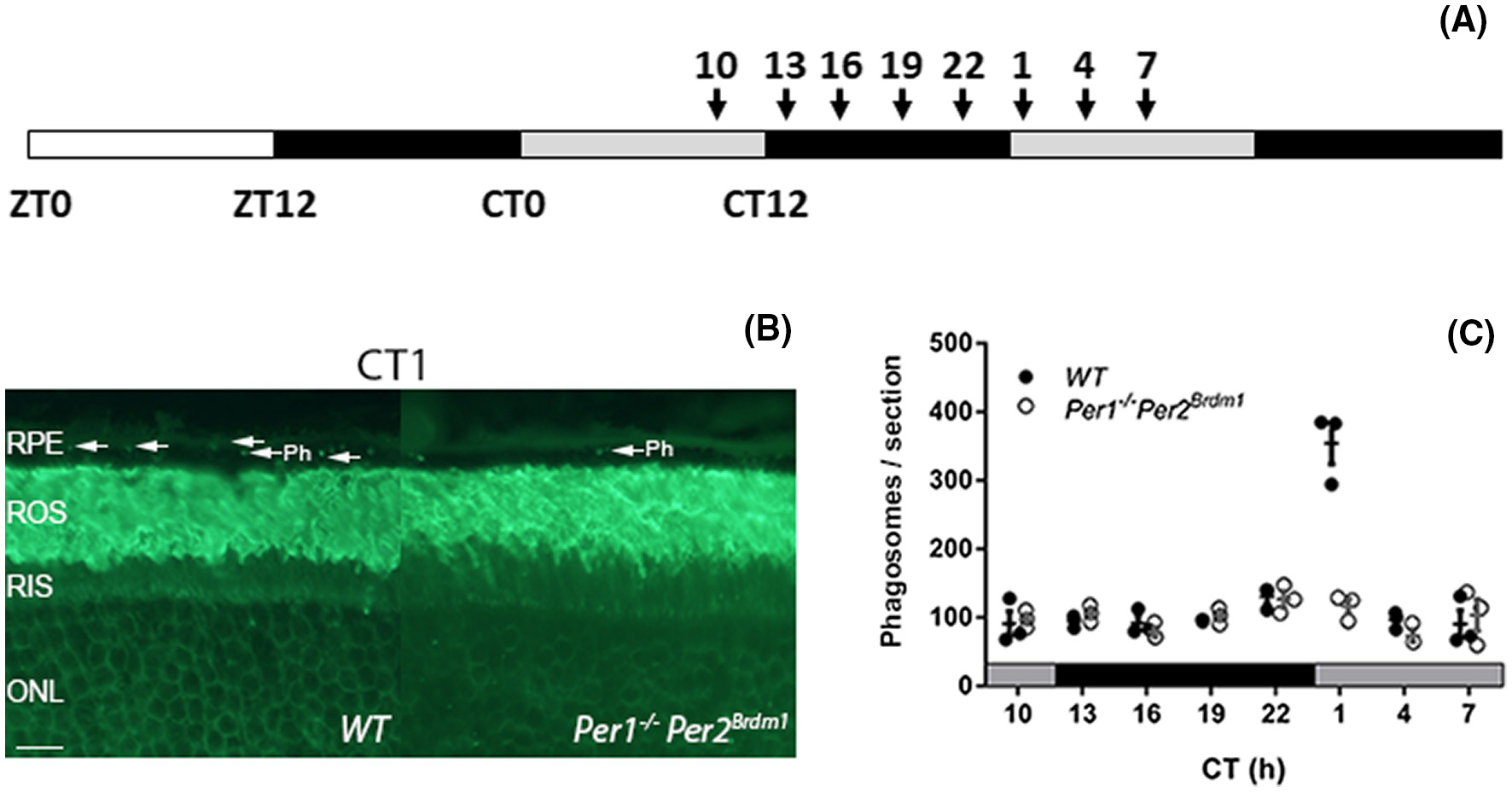 |
Mice lacking Per1 and Per2 show an impaired peak in POS phagocytosis. A, WT and Per1−/−Per2Brdm1 mice maintained under 12 hours light (white bar)-dark (black bar) conditions were placed under constant darkness (DD, grey-black bars) and sacrificed at time points indicated by arrows. B, Representative image of Rho-4D2 stained phagosomes of WT and Per1−/−Per2Brdm1 retinas obtained at CT1 during the peak in phagocytosis in DD conditions. ONL, outer nuclear layer; Ph, phagosomes; RIS, rod inner segments; ROS, rod outer segments; RPE, retinal pigment epithelium. The scale bar is 10 µm. C, Quantification of phagosomes in WT and Per1−/−Per2Brdm1 retinas under DD showed that Per1−/−Per2Brdm1 mice had no detectable peak in ROS phagocytosis. N = 3/genotype/time point. Graphs show mean ± SEM and values from individual samples are shown as dots |
Further analysis on the pathway level suggested a group of interacting genes that potentially drive the POS phagocytosis in the RPE. Per1 and Per2 are thought to be necessary clock components driving POS pghagocytosis in a process that is transcriptionally driven by the RPE.
To characterize the potential link between the circadian clock and the peak in POS phagocytosis time-affected transcriptomes of the RPE and photoreceptors were characterized. WT and Per1−/−Per2Brdm1 mouse eyes kept in DD at 4 time points (CT19, 22, 1, and 10) were harvested and the RPE and photoreceptors from each eye were laser-capture microdissected and RNA sequencing was performed. The RNASeq analysis was performed by the Bioinformatics Laboratory (Aldo Jongejan & Perry Moerland) using the limma-voom pipeline and visualized using their in-house developed R/Shiny application. Performing pairwise comparisons between the different timepoints, large differences in the number of differentially expressed genes were found in the RPE and not in the photoreceptors, indeed suggesting the RPE to drive the phagocytosis.
To further investigate this, potential gene pathways and protein-protein interactions were inferred using the GO and STRING databases. RPE genes potentially involved in initiating the peak in POS phagocytosis were thus identified.
| Identification of potential phagocytic pathways in RPE. A, A comparison of WT and Per1−/−Per2 Brdm1 (KO) RPE transcriptomes within each time point revealed that most genes were differentially expressed during the peak phagocytosis time point—CT1. Red numbers represent the number of upregulated differentially expressed genes, whereas blue ones are downregulated. B, Selection strategy for compiling the list of genes in the RPE possibly implicated in regulating POS phagocytosis. Signature rod and cone genes48 and the Gene Ontology term “Photoreceptor Outer Segment” were used to remove photoreceptor genes from the list of genes that potentially regulate POS phagocytosis. C, Functional enrichment analysis using g:Profiler showed that these genes are enriched in neurotransmission-related pathways from the WP, Reactome, KEGG databases. The orange line represents the significant cut-off (adjusted P < .05). D, STRING network analysis of protein functional associations of products of RPE genes implicated in initiating phagocytosis. Nodes represent protein products (n = 57). Disconnected nodes are not shown. Edges represent protein functional associations. Interaction confidence scores range 0.25-0.99 | 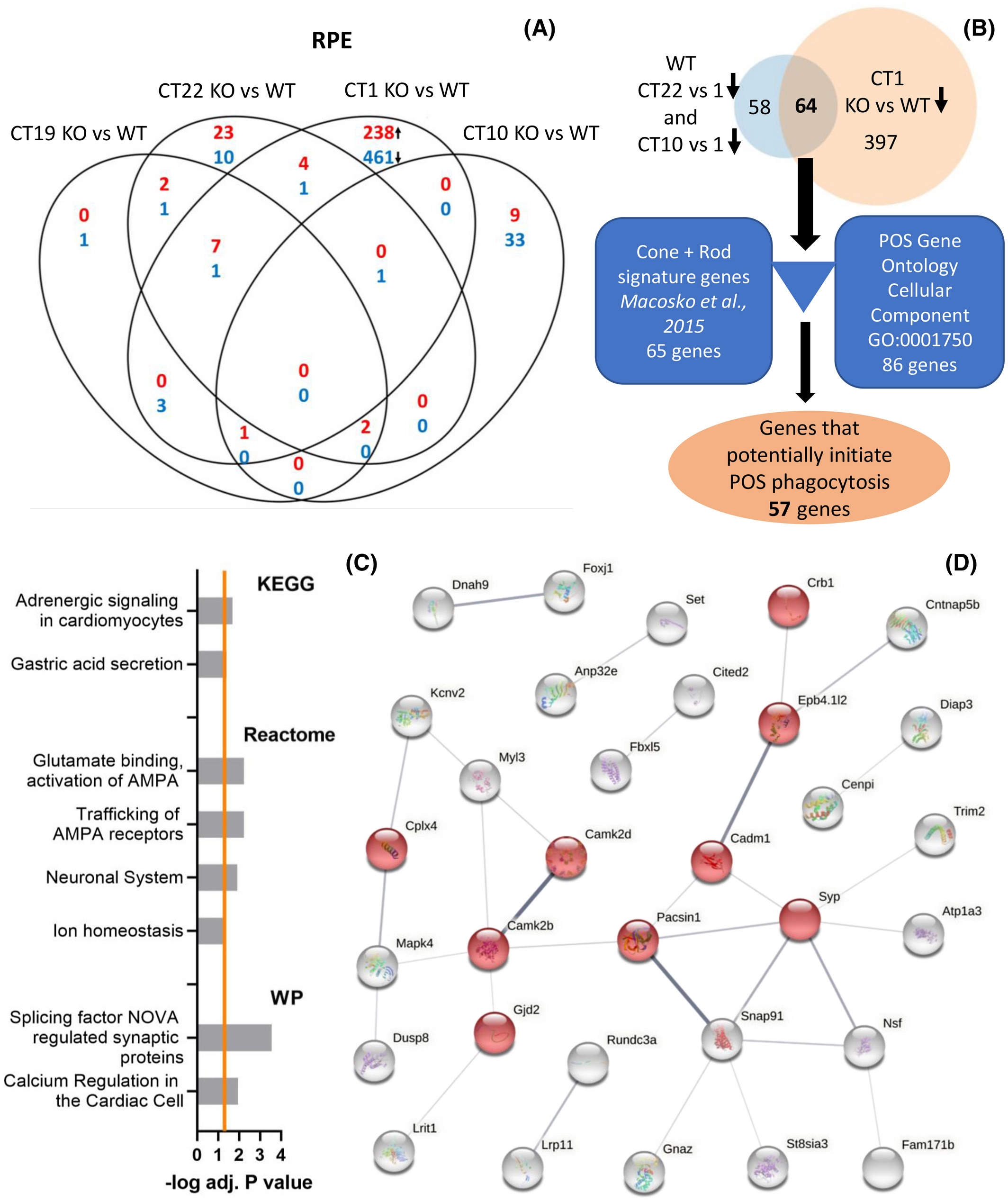 |
Core circadian clock genes Per1 and Per2 regulate the rhythm in photoreceptor outer segment phagocytosis
Nemanja Milicevic, Ouafa Ait-Hmyed Hakkari, Udita Bagchi, Cristina Sandu, Aldo Jongejan, Perry D. Moerland, Jacoline B. ten Brink, David Hicks, Arthur A. Bergen, Marie-Paule Felder-Schmittbuhl
FASEB J., 2021, 35(7) e21722